The Human Eye
The human eye or eyeball is the organ of sight. It is shaped like a slightly flattened sphere and the way it works is very complex, often compared to the workings of a camera.

Eyebrows
The eyebrows are made up of the set of small hairs located on or above the upper edge of the eye socket. The main function of the eyebrows is to prevent impurities or body fluids (such as sweat) trickling into the eye from the forehead.
Related Pathologies
BlepharitisConjunctiva
The conjunctiva is a thin, transparent and vascularised membrane. It covers the inner surface of the eyelids and then folds over to cover the front part of the eyeball (the sclera) as far as the edge of the cornea. The conjunctiva is the first site of immune response when the eye comes into contact with infectious or irritant agents.
Related Pathologies
PtosiEctropionEntropionHypermetropiaDry-eyeBacterial conjunctivitisAllergic conjunctivitisKeratitisTear duct disordersMyopiaPresbyopiaAstigmatismSclera
The sclera is usually identified as the “white of the eye”. This is a rather rigid fibrous tissue with a primarily structural function, but it also protects the inner contents of the eye from trauma.
Related Pathologies
PtosiEctropionEntropionHypermetropiaDry-eyeBacterial conjunctivitisAllergic conjunctivitisKeratitisTear duct disordersMyopiaPresbyopiaAstigmatismChoroid
The choroid is a membrane rich in blood vessels that surrounds the eye and is located between the sclera and the retina. Its main function is to nourish the outer layers of the retina.
Related Pathologies
UveitisRetina
The retina is a light-sensitive nerve tissue that lines the inner surface of the posterior segment of the eyeball. Its main function is to perceive light stimuli via photoreceptors (called cones and rods) and convert them into nerve impulses to be sent to the brain via the optic nerve.
Related Pathologies
Diabetic retinopathyRetinal oedemaAge-related macular degeneration (AMD)Optic nerve
The optic nerve is the first part of the optic pathway, i.e. the route that a visual stimulus takes from the retina to the brain. The optic nerve is in fact made up of the long terminals of the ganglion cells, cells that are located in the retina in contact with the photoreceptors, from which they collect nerve impulses that they carry to the brain to be processed as images.
Related Pathologies
GlaucomaVitreous humor
The vitreous humour (or vitreous body) is a transparent gelatinous tissue, 99% water, which fills the eyeball cavity between the lens and the retina. It performs several functions: an optical one, because with its lens effect it plays a role in focusing; a structural one, as it gives volume and stability to the eyeball; and a trophic one, because it acts as a conduit and filter for nutrients to pass through.
Related Pathologies
Posterior vitreous detachmentLens
The crystalline lens is a true transparent “lens”, positioned behind the iris and in front of the vitreous body. It allows objects to be focused on the retina, changing its shape and curvature, depending on their distance through an automatic process called accommodation. Its nutrition is provided by the aqueous humour since there are no blood or lymph vessels in the lens.
Related Pathologies
CataractsCiliary body
The ciliary body is a ring-shaped structure with a triangular cross-section located between the iris and the choroid, together with which it forms the uvea. Its muscular component (the ciliary muscle) contributes to the focusing of images through its connections to the lens, while a series of small offshoots (called ciliary processes) are responsible for the production of aqueous humour.
Related Pathologies
UveitisEyelid
The eyelids are the two movable structures, consisting of skin on the outside and mucous membrane on the inside, that cover the otherwise exposed front part of the eyeball. The main function of the eyelids is to protect the eyes from light and any foreign particles. In addition, their movement (blinking) distributes tears evenly over the ocular surface to always keep it moist.
Related Pathologies
StyeChalazionLacrimal apparatus
The lacrimal apparatus consists of the lacrimal glands and the lacrimal pathways, which are respectively responsible for the production and outflow of tears, as well as the actual tears, whose main function is to keep the surface of the eye constantly moist and clean, thanks to their turnover.
Related Pathologies
PtosiEctropionEntropionHypermetropiaDry-eyeBacterial conjunctivitisAllergic conjunctivitisKeratitisTear duct disordersMyopiaPresbyopiaAstigmatismEyelashes
Eyelashes are a set of small curved hairs on the edges of the eyelids. The main function of the eyelashes is to protect the eye against the entry of impurities and small foreign bodies (dust, insects, etc.).
Related Pathologies
BlepharitisAqueous humour
Aqueous humour is a colourless saline liquid produced by the ciliary body, which fills the space between the lens and the cornea. It performs several functions: an optical one, because with its lens effect it plays a role in focusing; a structural one, as it gives volume to the eyeball; and a trophic one, as it provides nourishment to the structures that it moistens.
Related Pathologies
Ocular hypertensionIris
The iris is located behind the cornea and in front of the lens, and is the “coloured” part of the eye. At the centre of the iris there is the pupil, the “hole” that widens and narrows under the influence of light to regulate the entry of light rays into the eye.
Related Pathologies
UveitisCornea
The cornea is a transparent, dome-shaped structure made up of five layers and without blood vessels, covering the front surface of the eyeball. The cornea has its own dioptric power and, just like a lens, it allows light and images to pass through, helping to focus them on the retina.
Related Pathologies
PtosiEctropionEntropionHypermetropiaDry-eyeBacterial conjunctivitisAllergic conjunctivitisKeratitisTear duct disordersMyopiaPresbyopiaAstigmatismEyelid
The eyelids are the two movable structures, consisting of skin on the outside and mucous membrane on the inside, that cover the otherwise exposed front part of the eyeball. The main function of the eyelids is to protect the eyes from light and any foreign particles. In addition, their movement (blinking) distributes tears evenly over the ocular surface to always keep it moist.
Related Pathologies
StyeBlepharitisChalazionLacrimal Gland
The lacrimal apparatus consists of the lacrimal glands and the lacrimal pathways, which are respectively responsible for the production and outflow of tears, as well as the actual tears, whose main function is to keep the surface of the eye constantly moist and clean, thanks to their turnover.
Related Pathologies
PtosiEctropionEntropionHypermetropiaDry-eyeBacterial conjunctivitisAllergic conjunctivitisKeratitisTear duct disordersMyopiaPresbyopiaAstigmatismHow does it work?
The eye obtains information about its surroundings through lightsmall parts works in synergy with the others to visualise the world around us, transmitting messages to the brain, which is then able to interpret the various messages.
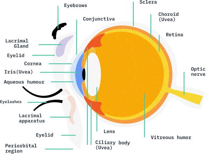
The light reaches the iris, which filters the intensity and helps the lens and the vitreous in the focusing process. This first process enables the retina to transform light into electrical signals, which are used to process images with the help of the optic nerve, the brain’s messenger.
Curious to know more about the anatomy of the eye?
Click on the + buttons to learn more